Tuesday, September 25, 2012

FOP Skeleton....

The Skeleton of Harry Eastlack

The skeleton of Harry Eastlack (1933 – 1973), a man who lived with FOP until he died just six days short of his fortieth birthday, is on display at The Mutter Museum of The College of Physicians in Philadelphia.
At age 5, he broke his left leg while playing with his sister. There were complications with the fracture, which did not set properly. The hip and knee stiffened and bone growths began to develop on the muscles of his thigh. Within some years the condition spread to other parts of his body, ossifying his tendons and muscle and fusing his joints. By his mid-20s his vertebrae had fused together. He died from pneumonia in November 1973, six days before his 40th birthday. At the time of his death his body had completely ossified; even his jaw locked up, leaving him able to move only his lips.


Late in his life, Harry Eastlack made the decision to bequeath his body to his physician who donated Harry's skeleton to The Mutter Museum so that physicians and scientists in future generations could study and learn about FOP. Unfortunately, when Harry was alive, he never met another person with FOP. Harry Eastlack's skeleton, one of the few existing in the world, has become a valuable asset to physicians and scientists studying Fibrodysplasia Ossificans Progressiva (FOP). 


The glass case in which Harry Eastlack's skeleton dwells has become a window into the medical mysteries and scientific challenges of FOP. It also exemplifies the harsh reality of FOP more than any chart, slide, or clinical description could accomplish.

Normal skeletons collapse into piles of loose bones when the connective tissues that join bones together in life are removed. To be displayed in human form, skeletons have to be re-articulated or pieced back together with fine wires and glue. As a result of the bridges of bone that formed from FOP flare-ups, Harry Eastlack's skeleton is almost completely fused into one piece, which proved little challenge for the articulator's craft.


Sheets of bone cover Harry Eastlack's back. Ribbons, sheets, and plates of bone lock his spine to his skull and his skull to his jaw. Additional ribbons and cordons of bone span from the spine to the limbs and immobilize the shoulders, elbows, hips, and knees. Thin stalagmites of bone launch themselves from his pelvis and thighs. His upper arms are welded to his breastbone by slender white bridges of bone that cross his immobilized rib cage.


This FOP skeleton, which stands as Harry Eastlack stood in life, is a constant reminder of how far scientists have come in the research of FOP, but it is also a constant reminder of how much further they need to go.
Although no member of the FOP research team at The University of Pennsylvania in Philadelphia ever knew Harry Eastlack during his lifetime, they have come to know Harry well posthumously through his remarkable bequest. Dr. Frederick Kaplan said that “Harry's skeleton is like The Sphinx. It stands silently and elegantly and reveals its secrets if you ask the right questions,”….“When we discover something important about FOP in the laboratory, we return to visit Harry's skeleton in order to confirm the reality of the discovery. At other times, we may discover something about Harry's skeleton that sends us running back to the laboratory to test a new hypothesis about FOP,” Kaplan added. “The gift that Harry has given to the FOP community is inestimable and his bequest has given additional meaning and depth to medical and scientific research well beyond the confines of his mortal existence. I never knew Harry in life,” added Kaplan, “But I bless his memory every time I visit the museum where his legacy continues to educate and inspire.”
Source: ifopa

Saturday, September 22, 2012

Inheritance


How do people inherit Fibrodysplasia Ossificans Progressiva (FOP)?

FOP is inherited in an autosomal dominant pattern, which means one copy of the mutated gene in each cell is sufficient to cause the disorder.
Most cases of Fibrodysplasia Ossificans Progressiva (FOP) result from new mutations in the gene. These cases occur in people with no history of the disorder in their family. In a small number of cases, an affected person has inherited the mutation from one affected parent.

Friday, September 21, 2012

How it is possible....


How are changes in the ACVR1 gene related to health conditions?

Now we all know that Fibrodysplasia Ossificans Progressiva (FOP) is caused by mutations in the ACVR1 gene. All individuals with a definite diagnosis of FOP have a mutation in which the protein building block (amino acid) histidine’ is substituted for the amino acid ‘arginine’ at position 206 of the ACVR1 protein (written as Arg206His or R206H). Researchers believe that under certain conditions this mutation may change the shape of the receptor. This shape change may disrupt the binding of an inhibitor protein such as FKBP12 or interfere with other mechanisms that control activation. As a result, the receptor may be constantly activated (constitutive activation) even in the absence of ligands. Constitutive activation of the receptor causes overgrowth of bone and cartilage and fusion of joints, resulting in the signs and symptoms of Fibrodysplasia Ossificans Progressiva (FOP).

Thursday, September 20, 2012

Function of ACVR1 Gene....


What is the normal function of the ACVR1 gene?

The ACVR1 gene provides instructions for making the activin receptor type I protein, which is a member of a protein family called bone morphogenetic protein (BMP) type I receptors. BMP receptors span the cell membrane, so that one end of the protein remains inside the cell and the other end projects from the outer surface of the cell. This arrangement allows receptors to receive signals from outside the cell and transmit them inside to affect cell development and function.
Activin receptor type I is found in many tissues of the body including skeletal muscle and cartilage. It helps to control the growth and development of the bones and muscles, including the gradual replacement of cartilage by bone (ossification). This process occurs in normal skeletal maturation from birth to young adulthood.
Activin receptor type I is normally activated at appropriate times by molecules called ligands. Activation may occur when these ligands, such as BMPs, bind to the receptor or to other proteins with which it forms a complex. A protein called FKBP12 can inhibit activin receptor type I by binding to the receptor and preventing inappropriate (leaky) activation in the absence of ligand.

Wednesday, September 19, 2012

ACVR1 gene.....


The official name of the ACVR1 gene...

The official name of this gene is activin A receptor, type I.
ACVR1 is the gene's official symbol.

Where the ACVR1 gene located...


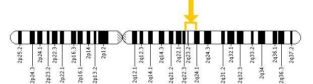
Cytogenetic Location:  2q23-q24

Molecular Location  : base pairs 158,592,957 to 158,732,373 ochromosome 2

The ACVR1 gene is located on the long (q) arm of chromosome 2 between positions 23 and 24.
More precisely, the ACVR1 gene is located from base pair 158,592,957 to base pair 158,732,373 on chromosome 2.


Tuesday, September 18, 2012

The Gene....


Which gene is related to Fibrodysplasia Ossificans Progressiva (FOP)?


Mutation in ACVR1 gene cause Fibrodysplasia Ossificans Progressiva (FOP).
The ACVR1 gene provides instructions for producing a member of a protein family called bone morphogenetic protein (BMP) type I receptors. The ACVR1 protein is found in many tissues of the body including skeletal muscle and cartilage. It helps to control the growth and development of the bones and muscles, including the gradual replacement of cartilage by bone (ossification) that occurs in normal skeletal maturation from birth to young adulthood.
Researchers believe that a mutation in the ACVR1 gene may change the shape of the receptor under certain conditions and disrupt mechanisms that control the receptor's activity. As a result, the receptor may be constantly turned on (constitutive activation). Constitutive activation of the receptor causes overgrowth of bone and cartilage and fusion of joints, resulting in the signs and symptoms of Fibrodysplasia Ossificans Progressiva (FOP).

Monday, September 17, 2012

The Discovery.....

The Gene Discovery
On 23rd April, 2006, after 15 years of painstaking research, the FOP research team at the University of Pennsylvania, School of Medicine and their international collaborators, pinpointed a single gene mutation—one out of 6 billion human genome—which is the culprit of such devastating bone growth in Fibrodysplasia Ossificans Progressiva (FOP).
Senior authors Eileen M.Shore, Ph.D. and Frederick S. Kaplan, MD, both from the Penn Department of Orthopaedic surgery and their international consortium of colleagues report their findings on April 23rd, 2006 in advance online edition of Nature Genetics”.
As per Kaplan “This discovery of the FOP gene is relevant to every condition that affects the formation of bone and every condition that affects the formation of the skeleton”.
Eileen Shore, a research associate professor of orthopaedic surgery at the University of Pennsylvania, has been studying the disease since 1991~ "I always had been interested in development on a cellular level" she says— "What changes a cell, or what regulates a cell to follow certain cell fate decisions? We usually think about development on an organism level, but it was more a question of what determines the personality of the cell?". 
In 2009, Shore and her team found some of the key biochemical steps that lead soft tissue cells to turn to bone—the results were published in the ''Journal of Clinical Investigation'' in November’09. 

Monday, September 10, 2012

Diagnosis


In most cases, an accurate diagnosis of Fibrodysplasia Ossificans Progressiva (FOP) can be made based on the patient's characteristic signs and symptoms of the disorder. Young children typically have malformed big toes and painful, tumour-like swellings on the head, neck, and/or back.


However, researchers believe that FOP is often misdiagnosed because it is a rare disease, affecting about one out of every two million people worldwide. Three most common misdiagnoses of FOP are Cancer, aggressive juvenile fibromatosis and progressive osseous heteroplasia. Experts estimate that the rate of misdiagnosis of FOP is arround 80% or higher.


Sunday, September 9, 2012

Complications


General: There are many complications associated with Fibrodysplasia Ossificans Progressiva (FOP) because the condition leads to progressive loss of mobility as more and more parts of the body become affected. Frequent illnesses or trauma to the body may speed up the disease progression.
Breathing problems: If extra bone replaces the tissues over the rib cage, the patient may not be able to expand their lungs normally. As a result, breathing may be difficult.
Difficulty speaking or eating: If extra bone develops in the jaw joints, a person may have difficulty speaking or eating. In some cases, difficulty eating may lead to malnutrition.
Limited mobility: As bone replaces muscles and connective tissues, the person with FOP typically experience limited mobility in the affected parts of their bodies. As a result day-to-day activities such as walking and getting dressed, may be challenging. Most FOP patients are bedridden by the time they reach 30 years of age.
Skin sores: People with FOP especially adults often develop pressure sores on their skin. Sore and broken skin may develop if extra bone puts pressure on the skin. Therefore, people with FOP should wear protective padding over bony bumps on their bodies and they are encouraged to change sitting positions frequently. People who sit for many hours during the day are encouraged to use well-padded seat cushions.
Skin sores may also develop if the skin becomes moist or sweaty in difficult-to-reach areas. Because people with FOP often have limited mobility of their joints, they may be unable to properly clean the skin. As a result, bacterial or fungal infections may develop on the skin.
Complications vary among patients with FOP, depending on which part of the body is affected.