Wednesday, August 29, 2012

History


The 1st case of Fibrodysplasia Ossificans Progressiva (FOP)

In 1692, French doctor Guy Patin was the first physician to record a case. The first thorough documentation of the disease was in 1740, when a London physician described an adolescent with large swellings of bone on his body in a letter to the Royal College of Physicians [source: IFOPA].
The best known FOP case is that of Harry Eastlack (1933–1973). His condition began to develop at the age of 10 and by the time of his death from pneumonia in November 1973, six days before his 40th birthday, his body had completely ossified, leaving him able to move only his lips.
Shortly before Eastlack's death, he made it known that he wanted to donate his body to science, in the hope that in death, he would be able to help find a cure for this little-understood and particularly devastating disease. Pursuant to his wishes, his preserved skeleton is now kept at the Mütter Museum in Philadelphia, and has proven to be an invaluable source of information in the study of FOP. There have approximately been 700 confirmed cases across the globe from an estimated 2500.

Tuesday, August 28, 2012

FOP Bone Vs. Normal Bone


Before moving to the FOP related facts and figures let’s try to understand FOP bones versus Normal bone.

FOP Bone versus Normal Bone

Bone is a living tissue. Each bone in our body is like an organ, made up of tissues and cartilage. FOP bone is just like normal bone — but in the wrong place.
Ossification (osteogenesis) is the process of new bone formation. There are two methods of bone formation:
ü Intramembranous 
ü Endochondral

Intramembranous bone formation is the simpler process, and it's responsible for forming a person's skull and lower jawbone. It's also determine how long bones like the ‘humerus’ and ‘femur’ grow in width.
Most bones in the body grow and heal after a break through endochondral bone formation. It's also controls how FOP bone grows. First, cartilage forms and then bone gradually takes the place of the cartilage.
Both kinds of ossification begin with mesenchyme. Mesenchyme is a connective tissue from which all other connective tissues come from. Mesenchymal cells can turn into different kinds of specialized cells which form tissues. The process of endochondral bone formation is as follows:
1.    Mesenchymal cells come together in the shape of the bone they will form. They turn into chondroblasts — cells that secrete cartilage matrix. A membrane called the perichondrium surrounds this cartilage.
2.    After the chondroblasts cover themselves in cartilage matrix, they turn into chondrocytes. The chondrocytes keep dividing while new chondroblasts continue to make cartilage matrix, causing the cartilage to grow.
3.    Some of the chondrocytes burst and others die. The bursting of the cells causes calcification or hardening of the cartilage. The dying cells make small spaces in the cartilage.
4.    nutrient artery enters the cartilage, triggering cells in the perichondrium to turn into osteoblasts. Osteoblasts are just like chondroblasts, but they secrete bone matrix instead of cartilage matrix. The osteoblasts start to secrete compact bone, and the perichondrium becomes the periosteum — the cover of the outside of the bone.
5.    Blood vessels grow into the cartilage and bring red bone marrow cells and other bone cells with them. The blood vessels stimulate a primary ossification center to grow — this is the place where bone tissue will begin to take the place of cartilage. Osteoblasts start covering the broken-down cartilage with bone matrix.
6.    Osteoclasts follow behind the osteoblasts and break down the spongy bone, making a cavity for red bone marrow to fill.
So, at this point, the long part of the bone, which started as cartilage, is compact bone with red bone marrow in the center. The endochondral bone formation finishes with the epiphyses, the ends of the bones. 
Secondary ossification centers develop to form bone, although unlike with the primary ossification center, spongy bone stays at the center of epiphyses instead of marrow.
This process uses undifferentiated cells or cells that can grow into any type of cell to make bones. The amazing thing about FOP is that the body convinces undifferentiated cells in tendons, ligaments and muscles to turn into something completely different. The body doesn't normally work this way. With FOP, ligaments and tendons and other connective tissues all go through this process of bone formation. It is normal bone, but in the wrong place at the wrong time — this is called heterotopic ossification.

So what happens with FOP?

A mutation in the gene encoding Activin receptor IA (ACVR1) tells the body to make an extra skeleton. This gene is responsible I controlling helps ‘bone morphogenetic proteins’ BMPs. In FOP, the gene is active without BMPs — operating like a leaky faucet. When BMPs are present, the faucet explodes with activity. This clue might someday help scientists figure out how to make extra bone for people who need it, like people with osteoporosis.


Monday, August 27, 2012

What is Fibrodysplasia Ossificans Progressiva (FOP)?


FOP !!! What is it?


Fibrodysplasia Ossificans Progressiva (FOP) is a rar autosomal dominant disorder with complete penetrance involving progressive ossification (where muscle tissue and connective tissue such as tendons and ligaments are gradually turned into bone) of  skeletal muscle, fascia, tendons and ligaments.


Any trauma to the muscles of an individual with fibrodysplasia ossificans progressive (FOP), such as a fall or invasive medical procedures, may trigger episodes of muscle swelling and inflammation (myositis) followed by more rapid ossification in the injured area. Flare-ups may also be caused by viral illnesses such as influenza.

The Nomenclature

FOP or Fibrodysplasia Ossificans Progressiva ~ fibro-dis-play-sha os-sih-fih-cans pro-gress-ev-a ~ means “Soft connective tissue that progressively turns into bone”


Sunday, August 26, 2012

The Mystery of Growth


We might not think about our bones very often unless we break one. When we break a bone, the bone heals itself and begins to regrow.
But, wait a minute!!!
What if our muscles, tendons and ligaments turned to bone?
What if we formed a second skeleton on top of the one we already have? 

That's what exactly happens with Fibrodysplasia Ossificans Progressiva (FOP).
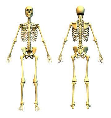
An FOP skeleton doesn't look like the ones we see at Halloween or the kind that hangs in an anatomy classroom. Instead of having lots of bones linked to one another with functioning joints, an FOP skeleton's bones fuse together, essentially forming a second skeleton out of the tendons, ligaments and muscles ~ a true metamorphosis. The skeleton is almost one solid piece, and sheets of bone exist where they should not.
The most common sign of FOP can be seen at birth: malformed big toes. Doctors aren't sure why this happens — it just appears as an early indicator of FOP. Aside from the malformed big toes, other initial signs of FOP usually show up in the early stage of life. One day, a large ‘lump’ suddenly begins to form on a child's body, usually in the ‘neck’ or ‘back area’. It can appear rapidly, often overnight and grows much faster than most tumours. It's warm to the touch, red and painful. A person's first reaction is often to assume it must be some sort of tumour — what if it's cancer? — but then the lump stops being painful, eventually gets smaller, and turns to bone — Normal Bone!!, but in the wrong place where the body neither needs it nor wants it.
These lumps are called Flare-Ups (a sudden burst of fire/light; here, a symptom of a disease) and they appear all through the life of a person with FOP. Doctors aren't always sure what triggers them, but they do know that any kind of injury, even a small one, can cause a flare-up. For someone with FOP, a fall is not just a fall and the typical bumps & bruises of daily life are a major threat to the mobility and independence. If a person with FOP bumps his elbow or knee, bone could begin to grow there and lock the arm or leg for the rest of the life.
In general, for FOP patients, extra bone formation almost always starts at the neck, spine and shoulders. Only then does it move to the other joints. Eventually, people with FOP will probably lose most of their mobility. Joints lock and bones can twist into odd positions. Some people with FOP develop scoliosis— their spine twists. Often, the jaw fuses together either spontaneously or as a result of an injection for dental work, which makes eating and brushing teeth extremely difficult.
The skeleton will fuse into one position and in that posture the person with FOP will stay in for the rest of his or her life.
Any attempt to remove the extra (heterotopic) bone only leads to extra bone formation.
Only 700 people worldwide are known to have FOP, which makes this disorder extremely rare [source: IFOPA].
The skeleton of Harry Eastlack, (1933 – 1973), suffered from FOP [from the collections of the Mütter Museum, College of Physicians of Philadelphia] © A.B. Shafritz et al., New Eng. J. Med. 1996, Massachusetts Medical Society.

Friday, August 24, 2012

Physiology of Nav1.7

Physiology of Nav1.7
In sensory neurons, multiple voltage-dependent sodium currents can be differentiated by their gating kinetics and voltage dependence and can also be defined by their sensitivity to the voltage gated sodium-channel blocker tetrodotoxin. The Nav1.7 channel produces a rapidly activating and inactivating current that is sensitive to submicromolar levels of tetrodotoxin. This is in contrast with Nav1.8, which is also present within DRG neurons but is fairly resistant to tetrodotoxin. Nav1.7 appears to be important in early phases of neuronal electrogenesis. Nav1.7 is characterized by slow transition of the channel into an inactive state when it is depolarized, even to a minor degree, a property that allows these channels to remain available for activation with small or slowly developing depolarizations, usually mimicked by electrophysiologists as ramp-like stimuli. Thus, Nav1.7 acts as a “threshold channel” that amplifies small, subtle depolarizations such as generator potentials, thereby bringing neurons to voltages that stimulate Nav1.8, which has a more depolarized activation threshold and which produces most of the transmembrane current responsible for the depolarizing phase of action potentials. In this regard, Nav1.7 is poised as a molecular gatekeeper of pain detection at peripheral nociceptors.

Thursday, August 23, 2012

Sodium channels

Sodium channels

Voltage-gated sodium channels play a critical role in the generation and conduction of action potentials — so important for electrical signalling by most excitable cells. Sodium channels are integral membrane proteins and are comprised of a large α subunit, which forms the voltage-sensitive and ion-selective pore, and smaller auxiliary β subunit(s) that can modulate the kinetics and voltage dependence of channel gating. Till 2007, 9 isoforms of the sodium-channel α subunit (Nav1.1– Nav1.9), each with a unique central and peripheral nervous system distribution had been identified. 4 closely related sodium channels (Nav 1.1, -1.2, -1.3, and -1.7) are encoded by a set of 4 genes (SCN1A, SCN2A, SCN3A, and SCN9A, respectively) located within a cluster on chromosome 2q24.3. Mutations in the genes encoding Nav1.1, -1.2, and -1.3 are responsible for a group of epilepsy syndromes with overlapping clinical characteristics but divergent clinical severity, mutation in the gene encoding Nav1.7 has a critical role in pain sensation.

Nav1.7 is encoded by SCN9A, a 113.5-kb gene comprising 26 exons (OMIM 603415). he encoded sodium channel is composed of 1977 amino acids organized into 4 domains, each with 6 transmembrane segments, and is predominantly expressed in the dorsal root ganglion (DRG) neurons and sympathetic ganglion neurons.

Immunohistochemical studies show that Nav1.7 is present at the distal ends of the wire-like projections of neurons known as neuritis, close to the impulse trigger zone where neuronal firing is initiated.


Interestingly, the large majority of DRG neurons that express Nav1.7 are pain sensing (nociceptive), suggesting a role for this sodium channel in the pathogenesis of pain. In addition to Nav1.7, Nav1.8 and Nav1.9 are also predominantly present in small nociceptive sensory neurons and the nerve fibres emanating from them.


Tuesday, August 21, 2012

Genetic Basis for Pain


The discovery of human genome allows comparing variations at the genetic level with inter-individual differences in pain thresholds and pain perception. Most studies in the past have focused on genetic polymorphisms that might be responsible for inter-individual differences in pain perception. Recently, genetic studies in families by James Cox and his team members, demonstrating recessively inherited CIP have identified nonsense mutations which result in truncation of the voltage-gated sodium channel type 1 x α subunit (SCN9A) — a 113.5 kb gene comprising 26 exons. The encoded sodium channel is composed of 1977 amino acids and is organised into 4 domains, each with 6 transmembrane segments. [Klugbauer et al. 1995]. The SCNA family of sodium channels (SCN1A-SCN11A) evolved from an archetypal potassium channel by quadruplicating, where 4 potassium subunits have to coalesce to form the functional potassium channel. SCN9A is predominantly expressed in the ‘dorsal root ganglion (DRG) neurons’ and sympathetic ganglion neurons. Functional studies, though performed for only some mutations to date, have shown that CIP associated mutations led to loss of function of Nav1.7.

Sunday, August 19, 2012

Body Parts Affected

What parts of the body does it affect?

It makes people with this condition feel no pain anywhere including the skin, deep tissues where special nociceptors called somatic pain are; and organs where the viceral pain nociceptors are. Because everything is surrounded by nerve endings to be managed by the brain, so everything feel pain, and is very dangerous in this condition that people don´t feel any pain, because they could die of an organ inflammation or some infection and they wouldn´t notice it.

Saturday, August 18, 2012

Cause


This autosomal recessive condition trait to chromosome 2q24.3. This region contains the gene SCN9A, encoding the α-subunit of the voltage gated sodium channel, Nav1.7, which is strongly expressed in nociceptive neurons. Sequence analysis of SCN9A in affected individuals revealed 3 distinct homozygous nonsense mutations:

ü W897X: Located in the P-loop of domain 2
ü 1767X: Located in the S2 segment of domain 2
ü S459X: Located in the linear region between 1 &2

This results in a truncated non-functional protein. Nav1.7 channels are expressed at high levels in nociceptive neurons of the ‘dorsal root ganglia’. As these channels are likely involved in the formulations and propagation of action potentials in such neurons, it is expected that loss of function mutation in SCN9A will lead to abolished nociceptive pain propagation.